Learn what a codon is, how codons work in protein synthesis, and their role in the genetic code. Complete guide with examples, types, and biological significance.
What is a Codon? Definition, Types, and How Codons Specify Amino Acids
A codon is a fundamental unit of the genetic code that serves as the basic "word" in the language of life. Understanding codons is essential for grasping how genetic information flows from DNA to proteins, making it one of the most important concepts in molecular biology.
Table of Contents
- Codon Definition
- Codon Structure
- Types of Codons
- Translation Process
- Genetic Code Table
- Biological Significance
- Usage & Optimization
- Important Examples
- Mutations & Effects
- Different Organisms
- Research Applications
- Analysis Tools
- Future Directions
- Common Misconceptions
Codon Definition: The Basics
Simple Definition
A codon is a sequence of three nucleotides (DNA or RNA bases) that corresponds to a specific amino acid or stop signal during protein synthesis.
Technical Definition
A codon is a triplet of nucleotides in messenger RNA (mRNA) that, when read by the ribosome during translation, specifies which amino acid should be added to the growing protein chain or signals the termination of protein synthesis.
Key Characteristics:
- Length: Always exactly 3 nucleotides
- Direction: Read in the 5' to 3' direction
- Non-overlapping: Each nucleotide belongs to only one codon
- Universal: The genetic code is nearly universal across all life forms
The Structure of a Codon
Nucleotide Composition
Each codon consists of three positions:
- First position (5' end): Usually the most constrained position
- Second position (middle): Often the most important for amino acid identity
- Third position (3' end): The wobble position with the most degeneracy
Example Codon: AUG
5' - A U G - 3'
     | | |
     1 2 3
- A (Adenine): First position
- U (Uracil): Second position
- G (Guanine): Third position
- Codes for: Methionine (Met)
- Special function: Start codon
Types of Codons
 Complete classification of all 64 codons in the genetic code
The genetic code contains exactly 64 possible codons (4³ = 64), which are classified into two main categories:
1. Sense Codons (61 total)
Sense codons are protein-coding codons that specify which amino acid should be added during translation:
Start Codon - The Translation Initiator
 AUG codon structure and its dual role in protein synthesis
-
AUG (Adenine-Uracil-Guanine): The universal start codon
- Amino acid: Methionine (Met)
- Function: Initiates protein synthesis in all organisms
- Location: Always the first codon in every protein-coding sequence
- Dual role: Also codes for internal methionine residues
-
Alternative start codons (organism-specific):
- GUG: Valine codon that can initiate translation in bacteria
- UUG: Leucine codon used as start in some prokaryotic genes
- Context-dependent: Require specific ribosome binding sites
Regular Amino Acid Codons (60 total)
Protein-coding codons that specify the 20 standard amino acids:
- 20 standard amino acids are encoded by these codons
- Codon degeneracy: Most amino acids have multiple codons (2-6 per amino acid)
- Wobble pairing: Third position often varies without changing the amino acid
- Codon families: Related codons often code for chemically similar amino acids
2. Stop Codons (3 total) - Translation Terminators
 The three stop codons that terminate protein synthesis
Stop codons (also called nonsense codons) signal the end of protein synthesis:
UAA - Ochre Stop Codon
- Most common: Frequently used stop codon across species
- Efficiency: Strong binding to release factors
- Recognition: Recognized by eRF1 in eukaryotes, RF1/RF2 in prokaryotes
UAG - Amber Stop Codon
- Research tool: Widely used in genetic studies
- Suppression: Can be read through by suppressor tRNAs
- Mutation: Often results from nonsense mutations
UGA - Opal Stop Codon
- Context-sensitive: Can code for selenocysteine in special contexts
- Variation: Codes for tryptophan in mitochondrial genetic code
- Suppression: Suppressible under specific conditions
Codon Degeneracy - The Genetic Code's Built-in Redundancy
Codon degeneracy (also called codon redundancy) means that most amino acids are specified by more than one codon. This redundancy provides evolutionary advantages and error tolerance.
Here is a breakdown of the degeneracy for each amino acid:
| Amino Acid | # of Codons | Codon Examples | Degeneracy Level |
|---|---|---|---|
| Methionine | 1 | AUG | No degeneracy |
| Tryptophan | 1 | UGG | No degeneracy |
| Phenylalanine | 2 | UUU, UUC | 2-fold |
| Tyrosine | 2 | UAU, UAC | 2-fold |
| Histidine | 2 | CAU, CAC | 2-fold |
| Glutamine | 2 | CAA, CAG | 2-fold |
| Asparagine | 2 | AAU, AAC | 2-fold |
| Lysine | 2 | AAA, AAG | 2-fold |
| Aspartic acid | 2 | GAU, GAC | 2-fold |
| Glutamic acid | 2 | GAA, GAG | 2-fold |
| Cysteine | 2 | UGU, UGC | 2-fold |
| Isoleucine | 3 | AUU, AUC, AUA | 3-fold |
| Threonine | 4 | ACU, ACC, ACA, ACG | 4-fold |
| Proline | 4 | CCU, CCC, CCA, CCG | 4-fold |
| Alanine | 4 | GCU, GCC, GCA, GCG | 4-fold |
| Glycine | 4 | GGU, GGC, GGA, GGG | 4-fold |
| Valine | 4 | GUU, GUC, GUA, GUG | 4-fold |
| Leucine | 6 | UUA, UUG, CUU, CUC, CUA, CUG | 6-fold |
| Serine | 6 | UCU, UCC, UCA, UCG, AGU, AGC | 6-fold |
| Arginine | 6 | CGU, CGC, CGA, CGG, AGA, AGG | 6-fold |
Key implications of codon degeneracy:
- Protection Against Mutations: A point mutation at the third position of a codon often does not change the amino acid, making the genetic code robust.
- Wobble Base Pairing: The third base of the codon can form non-standard base pairs with the anticodon of tRNA, allowing one tRNA to recognize multiple codons.
How Codons Work: The Translation Process
 Complete overview of how codons direct protein synthesis
Translation is the process where codons in mRNA are decoded to synthesize proteins. This fundamental biological process occurs in three distinct phases:
1. Initiation - Starting Protein Synthesis
 Ribosome assembly and start codon recognition
Start codon recognition and ribosome assembly:
Step 1: mRNA Binding
- Ribosome scans mRNA for the start codon (AUG)
- Shine-Dalgarno sequence (in prokaryotes) helps position the ribosome
- 5' cap recognition (in eukaryotes) initiates scanning
Step 2: First tRNA Recruitment
- Initiator tRNA (Met-tRNA) recognizes the start codon
- Anticodon (3'-CAU-5') pairs with codon (5'-AUG-3')
- Methionine is always the first amino acid in new proteins
Step 3: Translation Complex Assembly
- Large ribosomal subunit joins the small subunit
- P-site (peptidyl site) holds the initiator tRNA
- A-site (aminoacyl site) is ready for the next tRNA
2. Elongation - Building the Protein Chain
 Codon-by-codon protein synthesis cycle
Codon-by-codon translation cycle:
Step 1: Codon Recognition
- Next codon enters the ribosome's A-site
- Aminoacyl-tRNA with matching anticodon binds
- Codon-anticodon pairing ensures accuracy
- Proofreading mechanisms verify correct tRNA selection
Step 2: Peptide Bond Formation
- Peptidyl transferase (ribosomal RNA enzyme) catalyzes bond formation
- Amino acid from A-site tRNA joins the growing protein chain
- Peptide bond forms between adjacent amino acids
- Growing protein remains attached to A-site tRNA
Step 3: Translocation
- Ribosome moves one codon forward (5' to 3' direction)
- tRNA moves from A-site to P-site (carrying the growing protein)
- Empty tRNA moves from P-site to E-site (exit site)
- New codon enters the A-site for the next cycle
Key Features:
- Reading frame: Codons are read in groups of three without overlap
- Directionality: mRNA is read 5' to 3', protein grows N-terminus to C-terminus
- Speed: ~15-20 amino acids per second in eukaryotes
3. Termination - Completing Protein Synthesis
 Stop codon recognition and protein release
Stop codon recognition and protein release:
Step 1: Stop Codon Encounter
- Stop codon (UAA, UAG, or UGA) enters the A-site
- No tRNA exists with matching anticodon for stop codons
- Translation pauses as ribosome waits for binding
Step 2: Release Factor Binding
- Release factors (eRF1 in eukaryotes, RF1/RF2 in prokaryotes) recognize stop codons
- Class I release factors bind directly to stop codons
- Protein mimicry: Release factors structurally mimic tRNA
Step 3: Protein Release and Ribosome Recycling
- Peptidyl transferase hydrolyzes the bond between protein and tRNA
- Completed protein is released from the ribosome
- Ribosomal subunits dissociate and can be reused
- mRNA and tRNA are released for recycling
Quality Control:
- Nonsense-mediated decay: Eliminates mRNAs with premature stop codons
- No-go decay: Handles ribosomes stalled on problematic codons
- Ribosome rescue: Mechanisms to free stuck ribosomes
The Role of tRNA - The Codon Decoder
 Complete tRNA structure showing anticodon-codon pairing
Transfer RNA (tRNA) serves as the crucial molecular adapter that translates codons into amino acids. Each tRNA molecule is specifically designed to recognize one codon (or a small family of related codons) and deliver the corresponding amino acid.
tRNA Structure - A Molecular Precision Tool
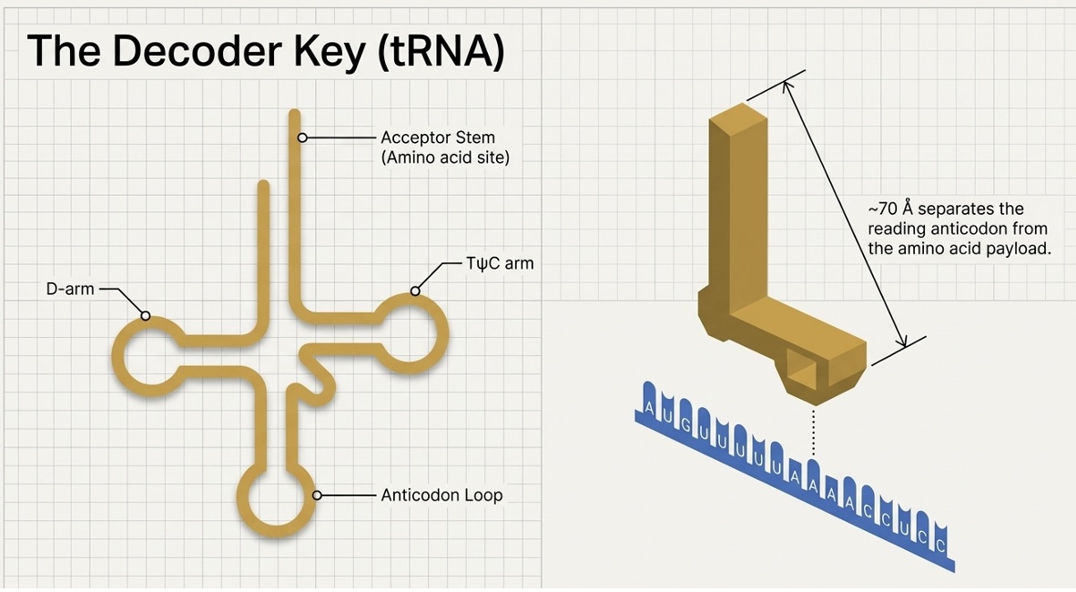 tRNA cloverleaf secondary structure and L-shaped tertiary structure
Secondary Structure (Cloverleaf Pattern):
- Acceptor stem: 3' end where amino acids attach (always ends in CCA)
- D arm: Contains dihydrouridine modifications for structural stability
- Anticodon arm: Contains the anticodon that pairs with codons
- TψC arm: Contains thymine and pseudouridine for ribosome binding
- Variable loop: Size varies between different tRNA species
Tertiary Structure (L-shaped):
- Anticodon loop: Positioned to interact with mRNA codons
- Amino acid attachment site: Located ~70 Å away from anticodon
- Elbow region: Flexible hinge allowing conformational changes
- Minor groove interactions: Precise codon recognition
How tRNA Decodes Codons - The Translation Mechanism
1. Aminoacylation (tRNA Charging):
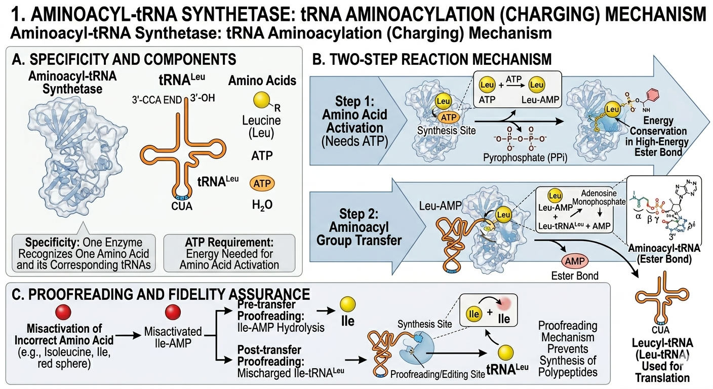 Aminoacyl-tRNA synthetase charging tRNA with amino acids
- Aminoacyl-tRNA synthetases attach specific amino acids to tRNAs
- Two-step mechanism: Amino acid activation + tRNA attachment
- Proofreading: Ensures correct amino acid-tRNA pairing
- ATP requirement: Energy needed for amino acid activation
- Specificity: Each synthetase recognizes only one amino acid and its corresponding tRNAs
2. Codon Recognition and Pairing:
 Detailed view of codon-anticodon base pairing in the ribosome
- Anticodon (3 nucleotides) pairs with codon (3 nucleotides)
- Watson-Crick base pairing: A-U and G-C pairs
- Wobble pairing: Flexible pairing at third position
- Induced fit: tRNA undergoes conformational changes upon binding
- Proofreading: Ribosome verifies correct codon-anticodon pairing
3. Amino Acid Delivery and Recycling:
- Peptide bond formation: Amino acid transfers to growing protein
- tRNA release: Deacylated tRNA exits the ribosome
- Recycling: tRNA can be recharged with new amino acid
- Quality control: Damaged tRNAs are degraded and replaced
Detailed Example: Phenylalanine tRNA in Action
 Step-by-step example of phenylalanine tRNA decoding UUU codon
Codon Recognition:
mRNA Codon:       5' - UUU - 3' (Phenylalanine codon)
tRNA Anticodon:   3' - AAA - 5' (Complementary sequence)
Base Pairing:     U-A, U-A, U-A (Watson-Crick pairs)
Amino Acid:       Phenylalanine (Phe)
Alternative Codon (Wobble Pairing):
mRNA Codon:       5' - UUC - 3' (Also codes for Phenylalanine)
tRNA Anticodon:   3' - AAG - 5' (Same tRNA can read both)
Base Pairing:     U-A, U-A, C-G (Third position wobble)
Amino Acid:       Phenylalanine (Phe)
Key Points:
- One tRNA can read multiple codons (wobble pairing)
- Codon degeneracy reduces the number of required tRNA species
- Accuracy: ~99.99% fidelity in codon recognition
- Speed: Each codon is decoded in ~20-80 milliseconds
The Genetic Code Table
 Complete genetic code table showing all 64 codons - Use our interactive codon table
Reading the Codon Table
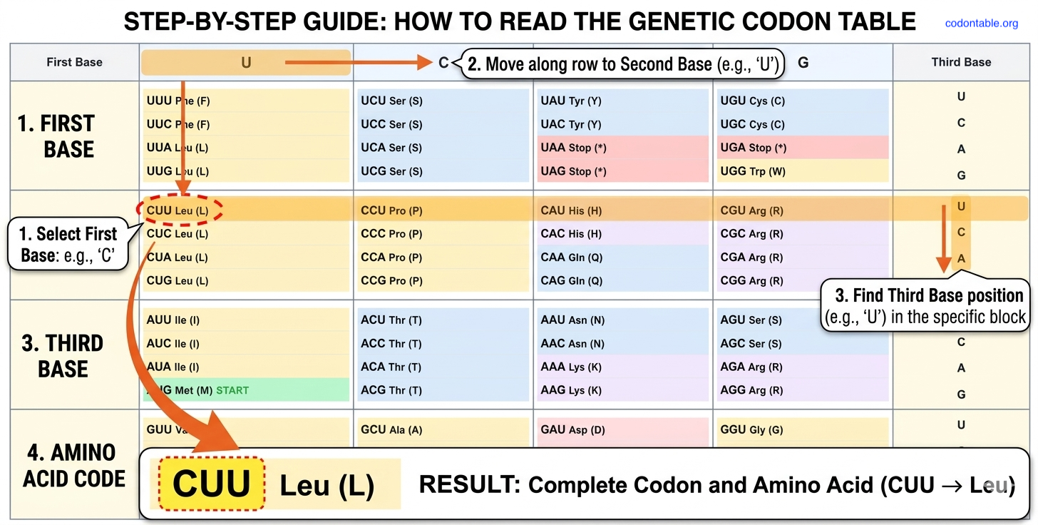 Step-by-step guide to reading the genetic code table
The codon table is organized as a systematic 4×4×4 grid that maps every possible three-nucleotide codon to its corresponding amino acid:
- First position (5' end): Determines the major codon family (U, C, A, G)
- Second position (middle): Most critical for amino acid specificity
- Third position (3' end): Shows "wobble" - often variable without changing the amino acid
Interactive Codon Reading Example
 Visual demonstration of how codons specify amino acids
Let's trace how codons work with a practical example:
UUU Codon Family Analysis:
5' → 3' Direction
┌─────┬─────┬─────┬──────────────────┐
│  U  │  U  │  U  │ → Phenylalanine  │
│  U  │  U  │  C  │ → Phenylalanine  │
│  U  │  U  │  A  │ → Leucine        │
│  U  │  U  │  G  │ → Leucine        │
└─────┴─────┴─────┴──────────────────┘
Key Observations:
- UUU and UUC: Both code for Phenylalanine (Phe) - demonstrating codon degeneracy
- UUA and UUG: Both code for Leucine (Leu) - showing wobble base pairing
- Pattern: The first two positions (UU) create a codon family, while the third position varies
Real-World Translation Example:
mRNA sequence: 5'-AUG UUU CCA UAG-3'
                  ↓   ↓   ↓   ↓
Amino acids:     Met Phe Pro Stop
Protein:         [Start]-Phe-Pro-[End]
Translation Process:
- AUG: Start codon → Methionine (protein synthesis begins)
- UUU: Codon → Phenylalanine (first amino acid added)
- CCA: Codon → Proline (second amino acid added)
- UAG: Stop codon → Translation terminates
Try our interactive codon table to practice reading codons yourself!
Wobble Base Pairing - The Third Position Flexibility
 Detailed mechanism of wobble pairing at the third codon position
Wobble base pairing is a crucial mechanism that allows flexibility in codon recognition, particularly at the third position. This flexibility reduces the number of tRNA species needed and provides evolutionary advantages.
The Wobble Position - Why the Third Position is Special
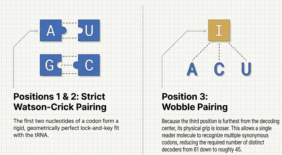 Importance of each codon position in amino acid specification
Position-Specific Constraints:
- First position: Highly conserved, critical for amino acid identity
- Second position: Most important for amino acid specification
- Third position: Most flexible, allows wobble pairing
- Structural basis: Third position is furthest from ribosome's decoding center
Standard vs. Wobble Base Pairs
Standard Watson-Crick Base Pairs:
- A-U (Adenine-Uracil): 2 hydrogen bonds
- G-C (Guanine-Cytosine): 3 hydrogen bonds
- Geometry: Perfect helical structure
- Stability: High binding affinity
Wobble Base Pairs:
- G-U (Guanine-Uracil): 2 hydrogen bonds, slight geometric distortion
- I-A (Inosine-Adenine): Modified purine allows flexible pairing
- I-C (Inosine-Cytosine): Inosine can adopt different conformations
- I-U (Inosine-Uracil): Broadest specificity wobble pair
Inosine - The Universal Wobble Base
Inosine structure and its ability to pair with multiple bases
Inosine Properties:
- Chemical structure: Hypoxanthine base (deaminated adenine)
- Pairing flexibility: Can pair with A, C, and U
- Evolutionary advantage: Reduces tRNA gene number
- Occurrence: Found in ~25% of tRNA anticodons
Wobble Pairing Rules and Examples
Codon Family Analysis:
1. Four-Codon Families (Complete Wobble):
Valine Codons:    GUU, GUC, GUA, GUG
tRNA Anticodons:  3'-CAA-5' (reads GUU, GUC)
                  3'-CAI-5' (reads GUA, GUG)
Result: 2 tRNAs read 4 codons
2. Two-Codon Families (Partial Wobble):
Phenylalanine:    UUU, UUC
tRNA Anticodon:   3'-AAG-5' (G can pair with both U and C)
Result: 1 tRNA reads 2 codons
3. Six-Codon Families (Complex Wobble):
Leucine Codons:   UUA, UUG, CUU, CUC, CUA, CUG
tRNA Set 1:       3'-AAI-5' (reads UUA, UUG)
tRNA Set 2:       3'-GAG-5' (reads CUU, CUC)
tRNA Set 3:       3'-GAI-5' (reads CUA, CUG)
Result: 3 tRNAs read 6 codons
Biological Significance of Wobble Pairing
1. Evolutionary Advantages:
- Silent mutations: Third position changes often don't affect protein
- Genetic robustness: Reduces impact of DNA replication errors
- Codon optimization: Allows fine-tuning of translation efficiency
- Species adaptation: Different organisms can use different codon preferences
2. Translation Efficiency:
- Reduced tRNA diversity: ~45 tRNA species instead of 61
- Faster translation: Fewer tRNA competition events
- Resource conservation: Less cellular energy spent on tRNA synthesis
- Quality control: Maintains translation accuracy while allowing flexibility
3. Therapeutic Implications:
- Nonsense suppression: Wobble tRNAs can read stop codons
- Codon optimization: Improving recombinant protein expression
- Genetic code expansion: Engineering new codon-amino acid pairs
- Disease treatment: Correcting genetic defects through codon manipulation
Biological Significance of Codons
1. Information Storage
- Genetic information: Codons store the blueprint for proteins
- Sequence specificity: Order of codons determines protein sequence
- Evolutionary conservation: Critical codons are highly conserved
2. Error Tolerance
- Synonymous mutations: Changes that don't affect amino acid
- Conservative substitutions: Similar amino acids from related codons
- Robustness: Genetic code minimizes harmful mutations
3. Regulation
- Codon usage bias: Organisms prefer certain codons
- Translation speed: Rare codons slow translation
- Gene expression: Codon choice affects protein levels
Codon Usage and Optimization
Codon Usage Bias and Optimization - Species-Specific Preferences
 Codon usage bias comparison across different organisms
Codon usage bias refers to the non-random use of codons in different organisms. Even though multiple codons can code for the same amino acid, organisms show strong preferences for specific codons.
Why Codon Usage Bias Exists
1. tRNA Abundance:
- Highly expressed genes use codons matching abundant tRNAs
- Translation efficiency: Preferred codons translate faster
- Resource optimization: Cells produce more tRNAs for common codons
2. Evolutionary Pressure:
- Selection for efficiency: Fast-growing organisms favor efficient codons
- Mutational bias: DNA repair and replication errors create patterns
- GC content: Organism-specific nucleotide composition preferences
Organism-Specific Codon Preferences
E. coli (Prokaryotic Model):
 E. coli codon usage preferences for major amino acids
| Amino Acid | Preferred Codons | Avoided Codons | Usage Ratio |
|---|---|---|---|
| Leucine | CUG (39%), UUG (13%) | CUA (4%), UUA (13%) | 10:1 |
| Arginine | CGU (36%), CGC (36%) | AGA (2%), AGG (2%) | 18:1 |
| Serine | UCU (16%), UCC (16%) | UCG (16%), AGU (16%) | 1:1 |
| Phenylalanine | UUC (57%) | UUU (43%) | 1.3:1 |
| Tyrosine | UAC (59%) | UAU (41%) | 1.4:1 |
Human (Eukaryotic Model):
 Human codon usage preferences showing different patterns from E. coli
| Amino Acid | Preferred Codons | Avoided Codons | Usage Ratio |
|---|---|---|---|
| Leucine | CUG (40%), CUC (20%) | UUA (7%), UUG (13%) | 6:1 |
| Arginine | AGA (20%), AGG (20%) | CGU (8%), CGA (11%) | 2.5:1 |
| Serine | AGC (24%), UCC (22%) | UCG (4%), UCA (15%) | 6:1 |
| Phenylalanine | UUC (54%) | UUU (46%) | 1.2:1 |
| Tyrosine | UAC (56%) | UAU (44%) | 1.3:1 |
Yeast (S. cerevisiae):
| Amino Acid | Preferred Codons | Avoided Codons | Usage Ratio |
|---|---|---|---|
| Leucine | UUG (28%), CUC (14%) | CUA (14%), CUG (11%) | 2:1 |
| Arginine | AGA (48%), CGU (15%) | CGG (3%), CGA (3%) | 16:1 |
| Serine | UCC (24%), UCU (26%) | UCG (9%), UCA (19%) | 3:1 |
Codon Optimization Strategies
 Step-by-step codon optimization process for heterologous expression
Step 1: Target Organism Analysis
- Identify host organism (E. coli, yeast, mammalian cells, etc.)
- Obtain codon usage tables from databases (CodonW, HIVE-CUTs)
- Analyze highly expressed genes in the target organism
- Determine tRNA abundance patterns
Step 2: Sequence Analysis
- Calculate current codon usage in the gene of interest
- Identify problematic codons (rare, avoided, or inhibitory)
- Check for secondary structures that might affect translation
- Analyze GC content and adjust if necessary
Step 3: Optimization Implementation
- Replace rare codons with preferred alternatives
- Maintain amino acid sequence (synonymous substitutions only)
- Avoid creating new problems:
- Restriction sites: Don't create unwanted cleavage sites
- Repeat sequences: Avoid long homopolymer runs
- Secondary structures: Prevent strong hairpin formation
- Ribosome binding sites: Don't create internal Shine-Dalgarno sequences
Step 4: Validation and Testing
- Synthesize optimized gene or use site-directed mutagenesis
- Test expression levels compared to original sequence
- Analyze protein functionality to ensure proper folding
- Optimize further if needed based on results
Advanced Optimization Considerations
1. Context-Dependent Effects:
- Codon pairs: Adjacent codons can influence translation speed
- Local tRNA depletion: Clusters of rare codons cause ribosome stalling
- mRNA secondary structure: Codon choice affects RNA folding
2. Expression Level Tuning:
- Moderate optimization: Partial optimization for controlled expression
- Full optimization: Maximum expression for industrial applications
- Deoptimization: Intentionally using rare codons to reduce expression
3. Organism-Specific Tools:
- Codon Optimization Tool: Interactive optimization calculator
- Expression Predictor: Estimate protein yield
- Sequence Analyzer: Comprehensive sequence analysis
Examples of Important Codons
Start Codons
AUG (Methionine)
- Universal start: Used by most organisms
- Dual function: Also codes for internal methionines
- Recognition: Shine-Dalgarno sequence in prokaryotes
Alternative Starts
- GUG: Valine codon that can initiate translation
- UUG: Leucine codon used as start in some genes
- Context dependent: Requires proper ribosome binding sites
Stop Codons
UAG (Amber)
- Nonsense mutation: Creates premature stop
- Suppressor tRNAs: Can read through in special cases
- Research tool: Used in genetic studies
UAA (Ochre)
- Most common: Frequently used stop codon
- Efficient termination: Strong release factor binding
- Evolutionary preference: Selected for reliability
UGA (Opal)
- Context sensitive: Can code for selenocysteine
- Mitochondrial variation: Codes for tryptophan in mitochondria
- Suppression: Possible with modified tRNAs
Codon Mutations and Their Effects
Types of Mutations
1. Silent Mutations
Original: CUU (Leucine)
Mutated:  CUC (Leucine)
Effect:   No change in protein
2. Missense Mutations
Original: GAG (Glutamic acid)
Mutated:  GUG (Valine)
Effect:   Different amino acid (sickle cell anemia)
3. Nonsense Mutations
Original: CAG (Glutamine)
Mutated:  UAG (Stop)
Effect:   Premature termination
4. Frameshift Mutations
Original: AUG CUU GCA...
Mutated:  AUG UCU UGC... (deletion of C)
Effect:   All downstream codons changed
Disease Examples
Sickle Cell Anemia
- Normal: GAG (Glutamic acid)
- Mutant: GUG (Valine)
- Effect: Altered hemoglobin structure
Cystic Fibrosis
- Common mutation: ΔF508 (deletion of phenylalanine)
- Codon affected: UUU or UUC
- Effect: Protein misfolding
Codons in Different Organisms
Prokaryotes vs Eukaryotes
Prokaryotes (Bacteria)
- Direct translation: mRNA translated during transcription
- Polycistronic mRNA: Multiple genes on one mRNA
- Shine-Dalgarno: Ribosome binding sequence
Eukaryotes
- Nuclear processing: mRNA modified before translation
- Monocistronic mRNA: One gene per mRNA
- 5' cap and 3' poly-A: Translation signals
Organellar Codes
Mitochondrial Codons
- UGA: Tryptophan (not stop)
- AGA/AGG: Stop (not arginine)
- AUA: Methionine (not isoleucine)
Chloroplast Codons
- Mostly standard: Few variations
- Bacterial origin: Similar to prokaryotic code
Research Applications
1. Genetic Engineering
- Codon optimization: Improve protein expression
- Site-directed mutagenesis: Change specific codons
- Synthetic biology: Design artificial genetic circuits
2. Evolutionary Studies
- Phylogenetic analysis: Compare codon usage patterns
- Selection pressure: Identify conserved codons
- Molecular evolution: Study genetic code evolution
3. Medical Applications
- Disease diagnosis: Identify pathogenic mutations
- Gene therapy: Design therapeutic genes
- Pharmacogenomics: Understand drug responses
Tools for Codon Analysis
 Analyze codons with our sequence translation tool - Try it now
Online Resources
- Interactive Codon Table - Visual codon lookup and analysis
- DNA to Protein Converter - Convert sequences and analyze codons
- Codon Wheel - Circular visualization of genetic code
- Codon usage databases: Organism-specific preferences
- Optimization software: Improve codon usage
- Mutation analysis: Predict effects of changes
Laboratory Techniques
- DNA sequencing: Determine codon sequences
- Site-directed mutagenesis: Change specific codons
- Ribosome profiling: Study translation in real-time
- Mass spectrometry: Analyze resulting proteins
Future Directions
Expanded Genetic Codes
- Unnatural amino acids: Beyond the standard 20
- Quadruplet codons: Four-base genetic codes
- Orthogonal systems: Independent translation machinery
Synthetic Biology Applications
- Biocontainment: Organisms dependent on artificial codons
- Enhanced proteins: Novel amino acid properties
- Metabolic engineering: Optimized enzyme production
Common Misconceptions
Myth 1: "All codons are equally important"
Reality: Start and stop codons are critical; others vary in importance
Myth 2: "The genetic code is truly universal"
Reality: Variations exist in mitochondria, chloroplasts, and some organisms
Myth 3: "Codon changes always affect proteins"
Reality: Silent mutations don't change the amino acid sequence
Myth 4: "Rare codons are always bad"
Reality: Sometimes used for regulation or proper protein folding
Conclusion
Codons are the fundamental units that translate genetic information into proteins. Understanding what a codon is and how codons function is essential for:
Key Takeaways:
- Basic unit: Codons are three-nucleotide sequences that specify amino acids
- Universal language: Nearly all life uses the same genetic code
- Error tolerance: The code is optimized to minimize harmful mutations
- Practical importance: Critical for genetic engineering and medicine
Why Codons Matter:
- Protein synthesis: Direct the assembly of all proteins
- Evolution: Provide the mechanism for genetic variation
- Medicine: Mutations in codons cause genetic diseases
- Biotechnology: Enable genetic engineering and synthetic biology
Whether you're a student learning molecular biology, a researcher working with genes, or simply curious about how life works at the molecular level, understanding codons provides insight into one of biology's most fundamental processes.
Related Concepts and Resources
- Codon Table Guide - Complete reference for the genetic code
- Central Dogma - How DNA→RNA→Protein works
- Stop Codons - Translation termination signals
- Genetic Code Variations - Alternative codes in different organisms
- Amino Acid Codon Chart - Comprehensive amino acid reference
- Codon Wheel - Visual codon organization
- Anticodons: Complementary sequences on tRNA molecules
- Reading frames: Different ways to group nucleotides into codons
- Mutations: Changes in codon sequences and their effects
Interactive Tools
- Interactive Codon Table - Explore codons visually
- DNA to Protein Converter - See codons in action
- Codon Wheel Visualization - Alternative codon chart format
Use our interactive tools to see codons in action and deepen your understanding of the genetic code.